2811103525 6979422768 Λεβήνου 74, Ηράκλειο gorgoraptisp@gmail.com


Prostate biopsy


A prostate biopsy is an invasive test that involves taking small tissue samples from the prostate gland. It is carried out when, during the screening (digital examination, rectal ultrasound, elevated P.S.A, multiparametric MRI), prostate cancer is suspected. In order to determine the type and extent of the disease, tissue samples (biopsies) must be taken from the prostate. These samples will be sent to a special laboratory for microscopic examination. Depending on the result of the microscopic examination, the appropriate treatment will be chosen.
To date, there is no hematological, imaging or any other method, apart from biopsy, that can reliably diagnose prostate cancer.
The urologist is the right doctor to judge whether a patient needs to undergo a prostate biopsy. A biopsy is done when a blood test shows an increase in prostate-specific antigen (PSA) or when the findings of a digital exam raise the suspicion of malignancy. More specifically, a prostate biopsy is necessary in cases where the probability of finding prostate cancer is increased. Such situations are the following:
• Suspicious finding on digital examination (hardness, asymmetry), regardless of PSA value
• Raised PSA value
• A large increase in PSA compared to a previous test
• Suspicious finding on multiparametric MRI


 How is a prostate biopsy done?

The examination is performed under local anesthesia, which makes it a relatively painless method. During preparation for the biopsy, the patient lies on the surgical bed in a lateral or gynecological position. Then the skin in the area of the genitals and anus is cleaned with an antiseptic solution and a special gel is inserted into the rectum with a local anesthetic. The prostate is located with the help of a special ultrasound head through the rectum. Under continuous ultrasound guidance, suspicious areas in the prostate can be identified. To avoid local pain and for easier insertion into the rectum, the ultrasound head is covered with the special gel which also contains local anesthetic, while anesthetic is injected around the prostate with a thin needle to avoid pain. A mapped tissue sample is then taken from the prostate gland using a fine needle fitted to a special automatic biopsy gun.


For the initial diagnosis, 10-12 systematic biopsies are usually taken from the peripheral zone of the gland. In some cases such as e.g. in a repeat biopsy it is necessary to obtain an even greater number of tissue pieces.
A repeat biopsy of the prostate is indicated if the indications persist:
• abnormal PE
• increased PSA value
• histopathological findings indicative of early malignancy in the initial biopsy (ASAP, HG PIN)
• positive mpMRI


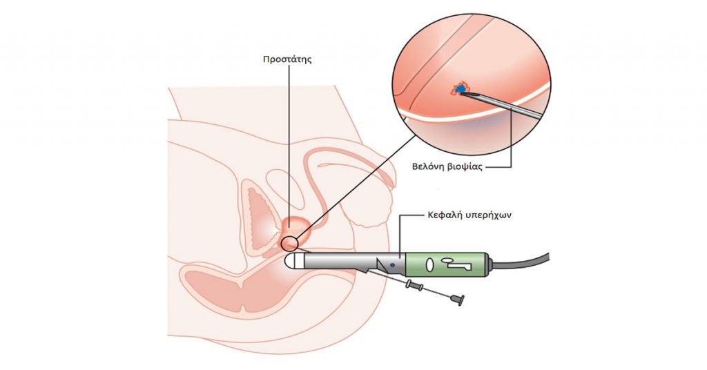
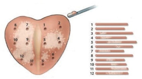
At the end of the examination, a gauze impregnated with local anesthetic and antiseptics is inserted into the rectum. The obtained tissue pieces from each area of the prostate are immediately placed separately in different vials with formalin solution and sent to the pathology laboratory for histological examination.
The opinion of the biopsy, done by the pathologist, who has specialized in the diagnosis of cancer and other pathological lesions of the tissues, usually shows one of the following conditions:
• Negative, i.e. without any evidence of cancer. Sometimes it can show evidence of prostatitis. But most of the time, evidence of hyperplasia and some other findings unrelated to cancer are described.
A negative biopsy does not completely rule out the presence of cancer, as a percentage of men with an initial negative biopsy will eventually be shown to have prostate cancer on a subsequent biopsy.
• Presence of so-called prostatic intraepithelial neoplasia (IN) or other precancerous lesions.
• Positive, i.e. definite presence of prostate cancer. The biopsy result should include the type of cancer, as well as parameters related to its extent (number of positive tissue sections, percentage of carcinoma per tissue section) as well as the Gleason Score (degree of aggressiveness of the disease).

The majority of examinees do not present any serious complication or disorder. Prophylactic antibiotics following the instructions given by the urologist helps to avoid possible infection. Blood in the urine may appear after the procedure, however after some hours and after a few emptyings of the bladder, it disappears. Rarely and in very serious cases, it is deemed necessary to insert a catheter to carry out washings. In patients with benign prostatic hyperplasia, urinary retention is likely to occur and catheter placement may be required.


1. Tell your doctor if:
• you have had any bleeding problems in the past
• you have had an allergy to any medicine
• you are taking medication for any reason
2. If you are taking anticoagulant drugs, which cause an increased tendency for bleeding, it is necessary to stop them for a certain period of time, which is determined by the attending physician. It is possible that you will need to take another anti-coagulant medicine, which causes less disruption of blood clotting (eg low molecular weight heparin).
3. Start the antibiotic 2 days before the biopsy and continue it after it for as long as the attending physician decides. In patients with an increased tendency to infections, antibiotic prophylaxis is of great importance. In patients with valvular disease or an artificial valve in the heart, it is necessary to take special antibiotics, which are determined by the cardiologist.
4. Because the biopsy is done through the intestine, you will need:
• the rectum has been emptied, otherwise a low enema is performed.
• the night before you should also avoid food (except for a yogurt or a soup).


What should you watch out for after the biopsy?

• Your urine may be pink or rarely red, or you may even urinate small clots of blood after the test or when straining during a bowel movement. This is normal. The presence of blood in the urine will stop on the same day or after two to three days.
• You should drink plenty of fluids, mostly water, to increase diuresis.
• You must avoid drinking alcohol for at least 2 days after the test.
• Take your antibiotics as prescribed by your doctor. Anticoagulant drugs should not be taken for a certain period of time after the biopsy, determined by your urologist.
• Avoid weight lifting and physical exertion for at least 7 days after the test.
• Blood may appear in the semen for several weeks after the biopsy. This is to be expected.
• Avoid any sexual intercourse for the first few days after the biopsy.

Contact your urologist immediately if you have:
• Fever (over 38°C) and chills.
• Urinary retention ie complete inability to urinate.
• Severe rectal bleeding or bleeding lasting more than 3 days.
• Severe hematuria or hematuria lasting more than 5 days or passing large blood clots in the urine.
• Intense pains.