2811103525 6979422768 Λεβήνου 74, Ηράκλειο gorgoraptisp@gmail.com


Lithiasis


What is urolithiasis?

Urinary lithiasis is the formation of a stone or stones in the drainage part of the urinary system (pelvic system of the kidney, ureter, bladder, urethra). It is the third most common urinary tract disease in humans, after urinary tract infections and prostate diseases. Depending on the location, we have lithiasis of the kidneys (nephrolithiasis), lithiasis of the ureters (the tubes that carry urine from the kidneys to the bladder), lithiasis of the bladder and finally lithiasis of the urethra. It is due to increased excretion of salts by the kidney, resulting in supersaturation of the urine, crystal formation and ultimately stone formation. Gallstones is a condition known since ancient times and is extremely common since 1 in 10 people will experience at least one episode of gallstones during their lifetime.

What are the properties of the stones
There are different types of stones that are determined based on their chemical composition.

- Calcium stones are the most common. They appear more frequently in men aged 30-40 years. Calcium forms complexes with other substances in the urine, such as oxalates and phosphorus, and eventually forms the stone (calcium oxalate and calcium phosphate).

- Uric acid stones are more common in men than women. The risk of their appearance increases in patients undergoing chemotherapy or suffering from gout
- Struvite stones (microbial stones) usually form in women suffering from persistent urinary tract infection. These stones can become very large in size.
- Cystine stones form in people suffering from cystinuria. This is an inherited disease that affects both sexes.

- Very rare forms of stones form in patients taking drugs such as indinavir and triamterene.


What are the risk factors?

The formation of stones in the urinary system is determined by endogenous (organic) and exogenous (environmental) factors. Usually, it is a combination of factors that contribute to the formation of stones.

Age: Stones most often appear between the ages of 20 and 50.
Gender: Men develop gallstones 2.5 times more often than women.
Family history-inheritance: About 25% of patients with lithiasis report having a first-degree relative with the same condition
Climatic conditions: People living in warm climates have an increased chance of developing stones (dehydration and supersaturation of urine)
Diet: Consuming an increased amount of animal protein and animal fat predisposes to stone formation, as does increased salt consumption. Also excessive consumption of foods rich in calcium (dairy) has been blamed for the development of lithiasis.
Obesity: Increased body weight predisposes to stone formation.
Fluid intake: Drinking little fluid, especially less than 1.5 liters per day, increases the chance of stone formation. Also, the trace element content of an area's drinking water can affect the disease.
Occupation: Office professionals have an increased incidence of nephrolithiasis compared to manual workers, a fact attributed to limited mobility, while stress also appears to increase the risk.
Medication: Specific antihypertensive medications, antacid medications, prolonged use of cortisone, and antiretroviral medications in HIV-predisposed patients to stone formation.
Anatomical or functional diseases of the urinary tract: Stenosis at some point of the ureter, stenosis at the pelivoureteric junction, spongiform kidney, calyceal diverticulum, vesicoureteral reflux, horseshoe kidney, ureterocele, nephrocalcinosis
Other systemic diseases: Hyperparathyroidism, gastrointestinal diseases (Crohn's disease, malabsorption diseases), sarcoidosis


What are the symptoms?

Many patients with urinary stones have no symptoms. The symptoms appear when a stone is found in the ureter, blocking the flow of urine from the kidneys. The most common symptom is renal colic, which is a very severe pain in the area of the kidneys, which may radiate to the abdomen, bladder or external genitalia. The pain usually comes on suddenly, is not relieved by changing position, and is often accompanied by a tendency to vomit (nausea) or even vomiting. Usually, there is hematuria which is microscopic and discovered in the urine and rarely macroscopic, which is seen during urination. Sometimes, when there is a urinary tract infection, lithiasis can present with symptoms such as chills, fever, fatigue, dysuria and constant kidney pain. Very often lithiasis is diagnosed incidentally in an imaging test done for another reason.


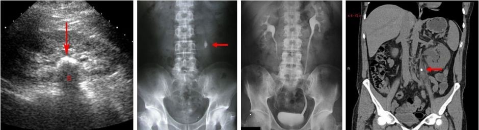
How is the diagnosis made?

The diagnosis of lithiasis is made by the history, the clinical examination and the necessary laboratory and imaging tests. The history and clinical examination raise the suspicion of lithiasis to the urologist, and the diagnostic tests are aimed at confirming the diagnosis, investigating the functionality and morphology of the urinary system, and detecting possible etiological factors responsible for the formation of stones. The tests used to investigate lithiasis are divided into imaging and laboratory tests. If you need to do some of these, your doctor will recommend it.


Visual inspection:

- Plain X-ray of kidneys, ureters and bladder (NOC): Detects about 70-80% of stones. However, there are stones, such as uric acid stones, which cannot be seen on X-ray NOK.
- Ultrasound of the urinary tract: Gives information about the existence of obstruction (hydronephrosis) and can detect stones that are inside the kidney or bladder but not in the ureter.
- Computed tomography without contrast agent: It is, today, the examination of choice in most urological centers for the investigation of lithiasis and at the same time a check is made of the anatomy of the kidney. It also provides information on the hardness of the stone.
- Intravenous urography or pyelography: It is an older method of imaging the urinary tract and is not often used.


Laboratory check:

- General urine: Usually, there are red blood cells in the urine and sometimes evidence of a urinary tract infection.
- Biochemical blood analysis: Measurement of urea and creatinine, uric acid and serum calcium.
- Parathormone: It is a hormone secreted by the parathyroid glands and is controlled in cases of increased calcium, as there may be hyperfunction of the glands as a cause of stone formation.
- 24-hour urine collection: The purpose is to determine the metabolic picture of the patient for the possible finding of some metabolic abnormality responsible for the production of stones.
- Analysis of the stone: It is done after the stone is found or removed in specialized laboratories, in order to find the type of stone and to take the appropriate measures to reduce the chances of recurrence and recurrence of lithiasis.

How is lithiasis treated?

Treatment of symptoms
In the phase of severe pain, renal colic, the aim is to relieve the patient of pain. Injectable drugs called non-steroidal anti-inflammatory drugs are given, which act as pain relievers and reduce local swelling. If the pain persists, stronger pain relievers, such as opioid analgesics, may be given. Then, to prevent colic episodes, non-steroidal anti-inflammatory drugs are given orally for 7 days and an α-adrenergic receptor blocker, which is usually taken by men for prostatic hyperplasia, can also be given. This treatment is considered to reduce the recurrence of colic and increase the possibility of spontaneous expulsion of the stone, since it causes damage to the ureter.

In a small percentage of patients, some immediate intervention will be needed when:
- pain persists despite medication
- the blockage threatens kidney function
- UTI coexists with fever.

In these cases, a ureteral catheter (pig-tail) may need to be inserted. Through the urethra with endoscopic instruments, the doctor reaches the bladder, finds the opening of the ureter and through it advances the catheter to the kidney. In cases where this is not possible, the tube is placed through the skin (percutaneous nephrostomy). If the conditions allow it, the removal of the stone can be done at the same time.


Pharmaceutical treatment

In some forms of stones, your doctor can prescribe you medicines, which aim either to reduce the size of the stone or to prevent its re-formation. Medicines differ depending on the chemical composition of the stone and whether there is any metabolic disease.


How are stones removed?

The removal of the stone depends on its size and location in the urinary system. As long as colic episodes are controlled with medication and for stones smaller than 6mm, up to 70% will pass the stone automatically within a 3-week waiting period. This interval is considered safe for maintaining the normal function of the kidney. In the remaining cases, invasive treatment will be needed to break up and remove the stone. Invasive methods include:
- Extracorporeal shock wave lithotripsy (ESWL): The stone is broken up with electromagnetic shock waves under fluoroscopy of the stone. It is usually very well tolerated by the patient and can be up to 90% effective. However, there are stones that cannot be broken by extracorporeal lithotripsy.
- Ureteroscopy and intracorporeal lithotripsy (URS): Used for stones located in the ureter. The ureteroscope through the urethra and bladder enters the ureter up to the stone, where special stone breakers, using a laser, break the stone, and its fragments are removed.
- Percutaneous nephrolithotripsy (PCNL): Used for large kidney stones (>2cm) or kidney stones that do not break with extracorporeal lithotripsy. Through a hole made from the skin to the kidney, the nephroscope is inserted, the stone is found and broken with special stone breakers, usually ultrasonic or ballistic.
- Open surgery: It has been limited today and is applied only in selected cases.


What is the necessary follow-up afterwards?

The probability of new stone formation one year after the initial stone episode is 10%, with the percentage increasing to 30-40% after 5 years and 50-60% in ten years. Therefore, it is necessary to monitor patients with lithiasis individually, depending on the individual's predisposition to create stones at intervals, which will be determined by the attending physician with a kidney-bladder ultrasound and plain X-ray of the kidneys, ureters and bladder (NIC).

Furthermore, it is necessary to take measures to avoid the recurrence of lithiasis. Your urologist will recommend that you:

Make sure you drink 2 to 2.5 liters every day
• Drink equally throughout the day
• Choose drinks with a neutral pH, specifically water
• Track how much you urinate. It should be about 2 to 2.5 liters a day
• Observe the color of your urine: it should be light
• Drink even more if you live in a hot climate or do a lot of physical exercise
Have a balanced and varied diet
• Eat plenty of vegetables, fiber and fruit (especially citrus fruits)
• Try to eat more low-oxalate foods, such as eggs, lentils, white rice, peeled apples, grapes, cauliflower, pumpkins, etc.
• Make sure your diet contains enough calcium (about 1,000 milligrams a day).
• Reduce the amount of salt in your diet (no more than 3 to 5 grams per day)
• Do not eat too much animal protein, especially meat from young animals. Instead, eat more plant-based protein, such as found in avocados, cauliflower or peas
• Maintain a healthy weight (your body mass index should be between 18-25kg/m2)